Plan B Aesthetics & Skincare
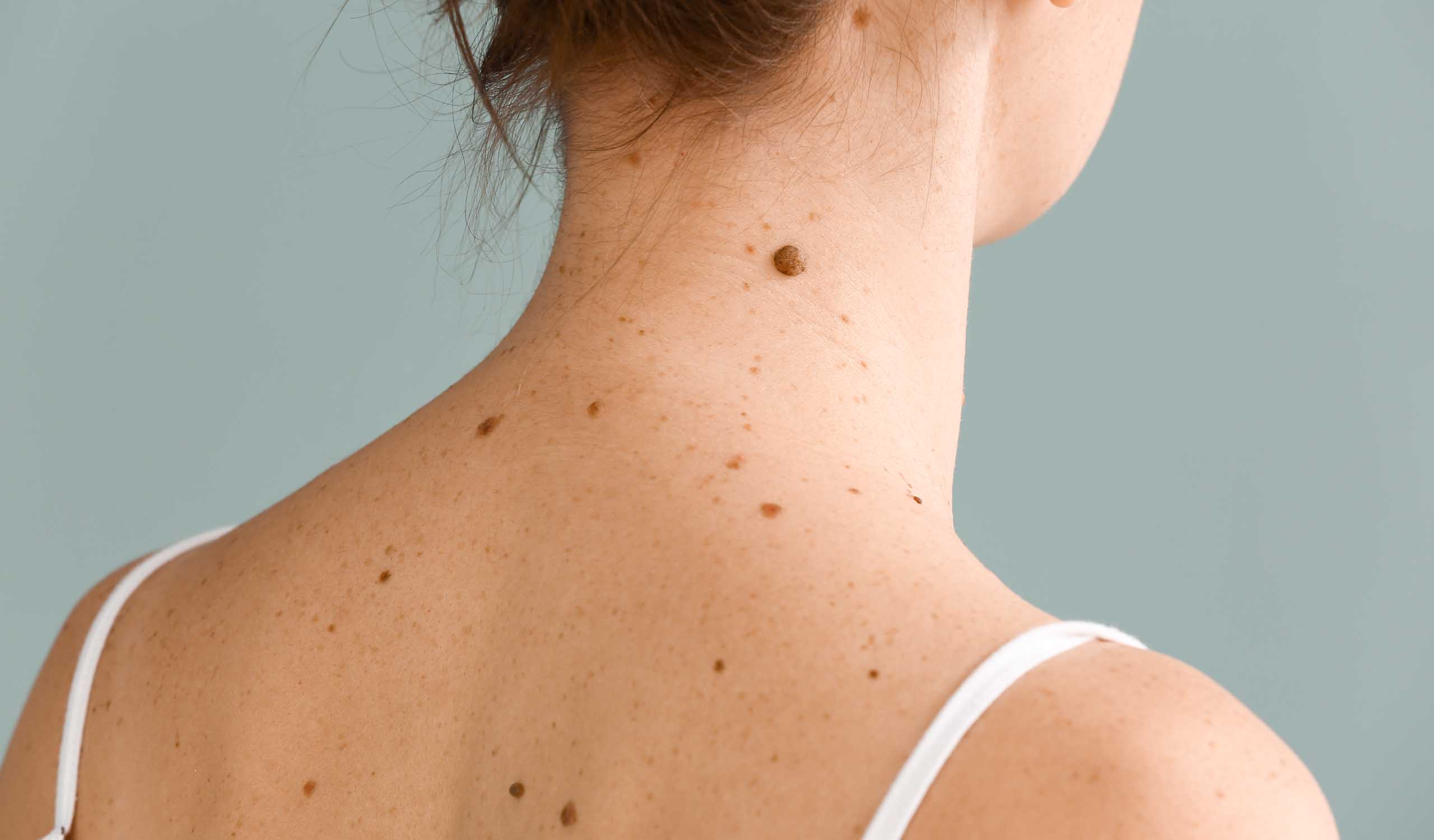
Identify and monitor potentially harmful moles
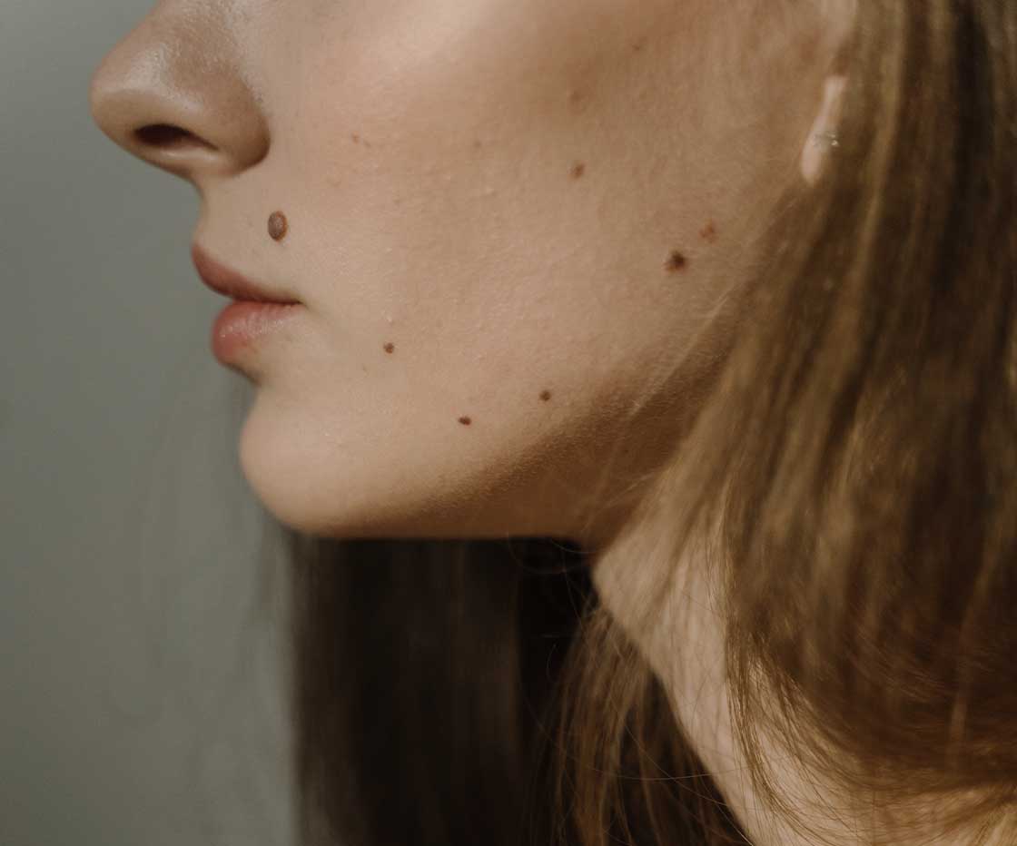
Map my mole facial mole mapping is a dermatological procedure that helps to distinguish between benign moles and potentially dangerous ones, that may be related to skin cancer. This procedure can eliminate the need for unnecessary biopsies and surgeries. By systematically documenting and monitoring moles on the face, and keeping a record, Dr Rachel can identify changes in size, shape, or colour that may indicate mole malignancy. This early detection is crucial for successful treatment and can significantly improve patient outcomes.
Your initial meeting with Dr Rachel will allow her to thoroughly evaluate your skin condition and medical history, ensuring that the treatment will be both effective and safe for you. During this time, Dr Rachel can identify any potential risks or concerns that may affect the outcome of the treatment. This personalised assessment helps tailor the procedure to your specific needs, maximising the chances of successful results. Ultimately, this thorough initial assessment sets the foundation for a successful map my mole treatment, aligning expectations and outcomes for optimal skin health.
Get in touchmap my mole
Why choose us?
As well as being highly trained and experienced in skin concerns, Dr Rachel is a dedicated member of the Clinical Medicine Aesthetics Council (CMAC), an organisation committed to raising safety standards in the aesthetic industry. By staying informed about the latest advancements and best practices, Dr Rachel ensures that her mole mapping treatments are conducted with the utmost care and precision.
By investing in top-tier map my mole equipment, Rachel ensures that each mole is thoroughly examined and documented, providing a reliable baseline for monitoring any changes over time. Her commitment to using only the best tools reflects her dedication to patient care and the importance she places on accurate diagnosis. Patients can feel confident that they are receiving the highest standard of care.
map my mole
Frequently asked questions
Mole mapping is a comprehensive method used to monitor and track changes in moles over time. When you undergo mole mapping, you can expect detailed images and records of your moles, which can be referenced in future examinations. This process helps in the early detection of any abnormalities or changes that might indicate skin conditions such as melanoma. The results from mole mapping will include high-resolution photographs of your skin, along with specific measurements and descriptions of each mole. Dr Rachel will analyse these images and compare them with previous records to identify any new or evolving moles. This meticulous documentation ensures any suspicious changes are promptly addressed, increasing the chances of successful treatment if needed.
Dr Rachel recommends that you have your moles mapped at least once a year. However, the frequency can vary depending on individual risk factors such as a personal or family history of skin cancer, the number and types of moles you have, and your skin type. Individuals with a higher risk may need to see Dr Rachel more frequently – potentially every 6 months.
Problematic moles include those that are atypical or dysplastic, meaning they have irregular shapes, uneven colours, or asymmetrical borders. Moles that change in size, shape, or colour over time are also concerning, as they may indicate malignant transformation. Additionally, moles that are larger than a pencil eraser or have a diameter greater than 6 millimetres should be monitored closely. It is also essential to pay attention to moles that itch, bleed, or become painful. People with a high number of moles or a family history of melanoma are at increased risk and should consider more regular mole mapping.
Dr Rachel will use a specialised camera and software to capture high-resolution images of each mole. There is no need for needles, incisions, or any form of physical discomfort. The most a patient might experience is the slight inconvenience of standing or positioning oneself in various ways to ensure all areas of the skin are adequately photographed.
During your mole mapping session, Dr Rachel will take high-resolution photographs of the specific areas of concern. These images are then analysed and stored for future comparisons. The primary goal of mole mapping is to track changes in the size, shape, colour, and texture of moles over time. By comparing current images with previous ones, Dr Rachel can identify any suspicious changes that might indicate malignancy.

your journey
1. Your Consultation
Informed consent is a critical part of the consultation process. Dr Rachel will explain the procedure, risks, benefits, and alternatives in detail, ensuring you understand and agree with the proposed plan of action. She will also review your family and medical history. This comprehensive approach helps provide peace of mind and supports effective skin health management.
Next step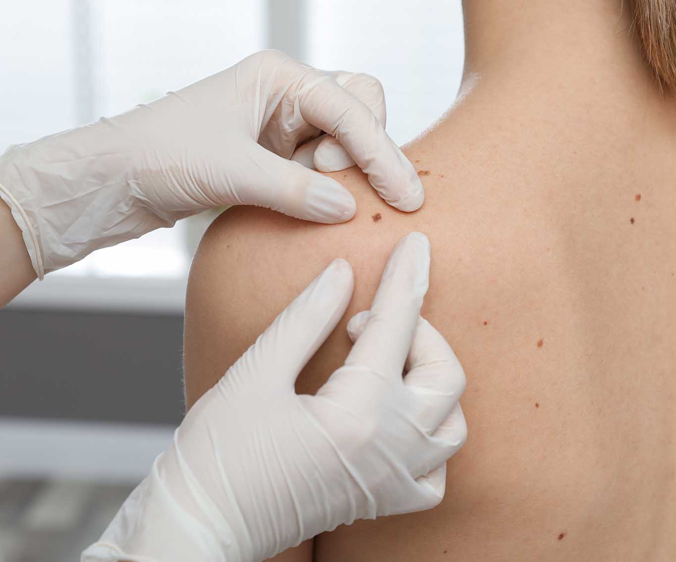
your journey
2. Your Treatment
During your map my mole procedure, a detailed record of all moles on your face is created using high-resolution imaging technology. This typically begins with the patient undergoing a full-face skin examination where Dr Rachel notes the size, shape, colour, and location of each mole. Following this examination, photographs are taken focusing on areas with moles. These images are stored digitally and can be compared with future images to identify any changes.
Next step
your journey
3. Your Aftercare
Patients should regularly check their skin for any changes in their moles, such as alterations in size, shape, colour, or texture, and report these changes promptly. Keeping a record of any new moles or changes can be beneficial for future consultations. Additionally, scheduling regular follow-up appointments with Dr Rachel is vital for ongoing monitoring and early detection of any potential issues.
Next step

